
This makes the assessment of diagnostic accuracy difficult due to the poor accuracy of chest radiographs. Most studies have compared ultrasound with chest radiograph findings. However, data on the sensitivity and specificity of ultrasound for PTB in children are still scanty. Diagnostic accuracy increases with user experience. Ultrasound abnormalities suggestive of PTB (consolidation, pleural effusion, or lymphadenopathy) may be found in 31 to 83% of confirmed cases with high inter-reader agreement. A major advantage of ultrasound is the ability to assess for features of extrapulmonary TB including ascites, hepatic micro abscesses, pericardial effusion or abdominal lymph nodes. Ultrasound allows trained clinicians to make prompt diagnostic and management decisions at the bedside.

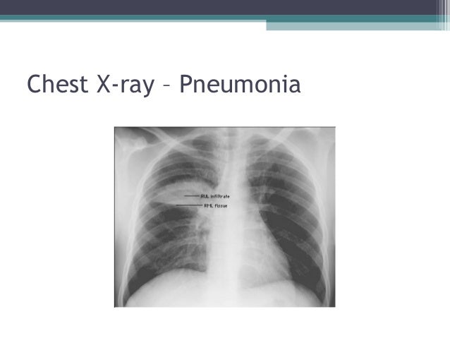
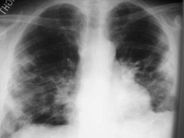
Longer scanning times (relative to other modalities)Ī chest ultrasound offers a portable, non-invasive, and real-time assessment of intrathoracic pathology without the use of ionizing radiation. Sensitivity/specificity comparable to CT (except small nodules/GGO)ĭifferentiate TB lymphadenopathy from reactive lymph nodes based on signal intensity and heterogeneity Ionizing radiation although low dose protocols now in useĬharacterization of lymph node morphology and enhancement May differentiate TB from non-TB lymphadenopathy Unable to assess pulmonary hila for lymphadenopathyĮarlier more sensitive detection of TB disease and complications compared with CXRĪbility to monitor disease complications and treatment response Sensitivity/specificity data for signs still scanty ĭetects mediastinal nodes or pleural effusion before CXR The detection of lymphadenopathy on chest radiographs has significant inter-observer variability (average weighted kappa of 0.33–0.36) and poor intra-observer agreement (average weighted kappa of 0.55). In another study, a moderate correlation between findings of lymphadenopathy on lateral chest radiograph and CT scan was shown, with precarinal lymph nodes associated with the highest sensitivity and specificity. One study found that a lateral radiograph in addition to a frontal image did not significantly improve the diagnostic yield, but increased cost and radiation exposure. The use of the lateral radiograph in improving sensitivity or specificity is controversial. Sensitivity of chest radiography for detecting lymphadenopathy, when compared against CT imaging, has been shown to be 67–74%, with a specificity of 39–59%. Despite the reliance on radiographs in the diagnosis of paediatric PTB, poor sensitivity, specificity, and wide inter observer agreement have been demonstrated. Parenchymal foci associated with lymphadenopathy (Ghon complex) are often small and difficult to identify on chest radiograph. Radiograph findings closely reflect the pathophysiology of the disease ( Table 1). Overview of Imaging Techniques AvailableĬhest radiographs are the primary radiologic investigation in children for diagnosis and assessment of PTB.


 0 kommentar(er)
